 This week in 1895, a monumental advancement in medical technology was made by accident in a German lab.
On November 8, 1895, German physicist Wilhelm Röntgen discovered X-rays while experimenting with vacuum tubes. Röntgen was investigating cathode rays with a fluorescent screen painted with barium platinocyanide and a Crookes tube which he had wrapped in black cardboard so the visible light from the tube wouldn’t interfere. He noticed a faint green glow from the screen, about 1 meter away. He realized some invisible rays coming from the tube were passing through the cardboard to make the screen glow.
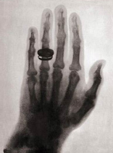
Röntgen immediately investigated these unknown rays. One week after his discovery, he took the first ever x-ray photograph of the human body. It was an picture of his wife’s hand showing her wedding ring and her bones.
 The first ever paper on x-rays was submitted to Würzburg’s Physical-Medical Society journal on December 28, 1895. Röntgen referred to the unknown type of radiation as “X” and the name stuck. Colleges suggested calling them Röntgen rays. In a few languages including German and Russian, the rays are still called “Röntgen rays.”
The development of X-rays was continued for use in the medical imaging. The first x-ray images were produced on glass plates. Eventually, glass plates were replaced by photographic film. The digital era has led to computerized and digital radiography.
From an accidental discovery in a German lab to digital radiography, X-rays have undergone quite a change. Still, they are one of the most important medical diagnostic tools in use.
